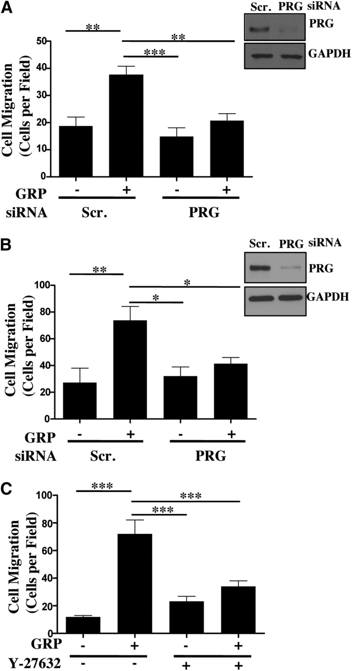
Fig. 6.
PRG-RhoA-ROCK axis mediates GRP-stimulated colon cancer cell migration. (A,B) Caco-2 (A) or HT-29 (B) cells transfected with scrambled or PRG siRNA for 48 hours. The transfected cells were serum-starved overnight and plated on the top chamber of Transwell insert at 5 × 10⁵ cells/well. The inserts were placed in 1% FBS containing media with or without 100 nM GRP (see Materials and Methods). Representative images of PRG knockdown in Caco-2 and HT-29 cells. Statistical analysis of cell migration of n = 3 repeated in duplicates. Shown are mean values ± S.E.M.; (*P < 0.05, **P < 0.01, ***P < 0.001). (C) Caco-2 cells were plated on the top of the Transwell inserts at 5 × 10⁵ cells/well in media with or without GRP along with Y-27632 (20 μM) (see Materials and Methods). Statistical analysis of cell migration of n = 3 repeated in duplicates. Shown are mean values ± S.E.M.; **P < 0.01, ***P < 0.001.

