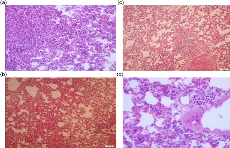
Fig. 1.
(a) Severe neutrophilic infiltration and increased alveolar wall thickness in the DIR group, HE×200. (b) Moderate neutrophilic infiltration and increased alveolar wall thickness in the DIRD group, HE×100. (c) Mild neutrophilic infiltration and increased alveolar wall thickness in the DC and D groups, HE×100. (d) Normal structure lung tissue parenchyma in the control group, HE×200.

