


Figure 2.
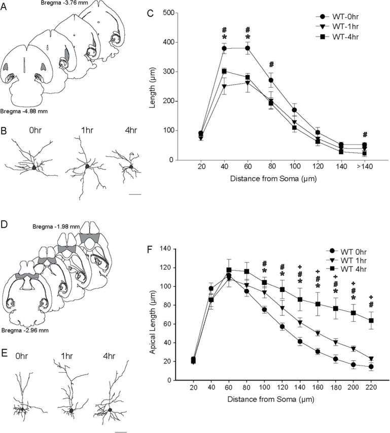
Acute restraint stress (ARS) remodels dendrites in the basolateral amygdala (BLA) and orbitofrontal cortex (OFC). (A) Schematic diagram of horizontal sections through the mouse brain. The shaded areas indicate portions of the BLA from which neurons were sampled. Coordinates indicate position in the dorsal-ventral axis relative to bregma (Paxinos and Franklin, 2001). (B) Reconstructions of representative pyramidal neurons from the BLAs in wildtype (WT) mice that were either unstressed (NRS; 0h) or underwent 1h or 4h of ARS. Neurons are near the mean for each group. Scale bar = 50 µm. (C) Mean length of BLA pyramidal neuron dendrites between 20-µm concentric spheres in mice that underwent 0h, 1h, or 4h of ARS. Dendritic retraction was evident immediately after either 1h or 4h of ARS. (D) Schematic diagram of horizontal sections through the mouse brain. The shaded areas indicate portions of the OFC from which neurons were sampled. (E) Reconstructions of representative pyramidal neurons from the OFCs in WT mice that underwent 0h, 1h, or 4h of ARS. (F) Mean length of OFC pyramidal neurons apical dendrites between 20-µm concentric spheres in WT mice that underwent 0h, 1h, or 4h of ARS. Either 1h or 4h ARS produced significant dendritic proliferation relative to NRS. For all graphs, *p < 0.05 for 0h vs. 1h; # p < 0.05 for 0h vs 4h; + p < 0.05 for 1h vs 4h.