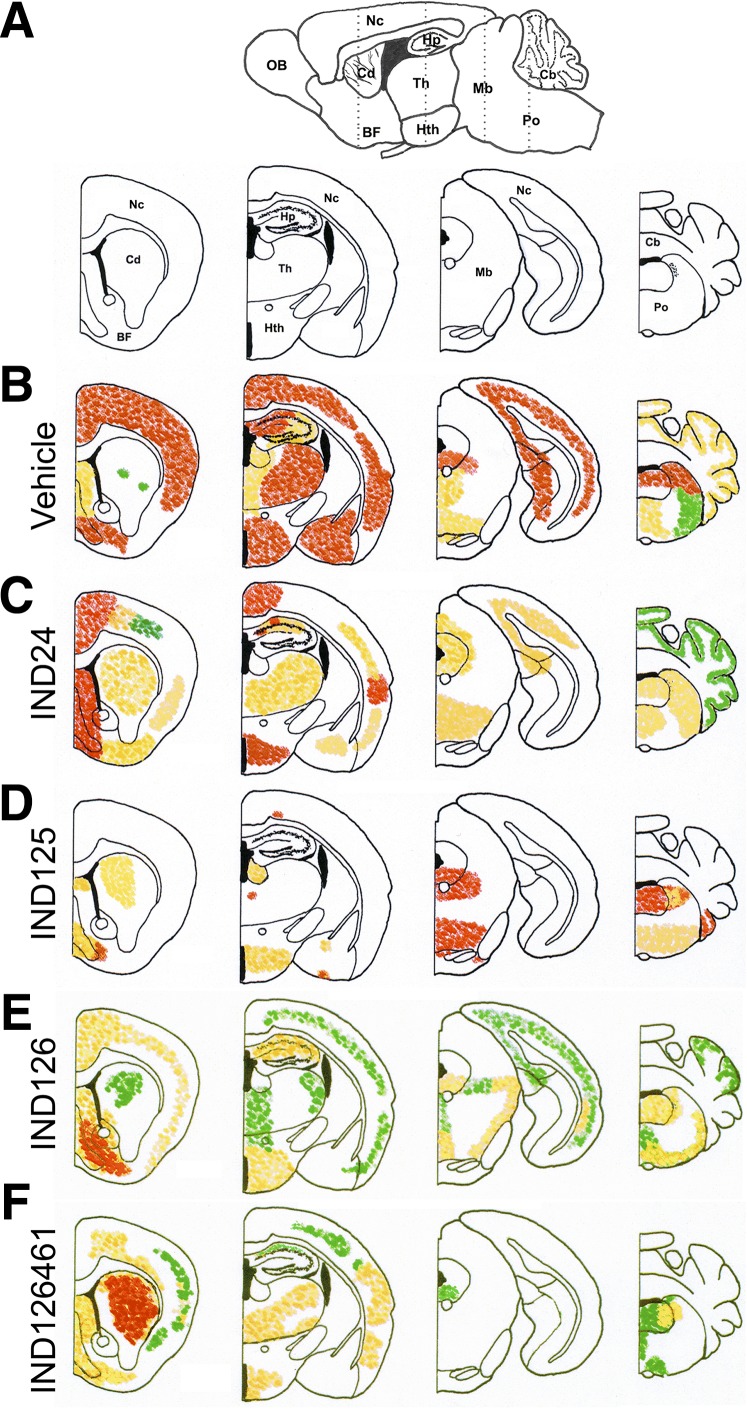
Fig. 4.
Neuropathological characterization of brains from Tg4053 mice treated with 2-AMTs. (A) Schematic sagittal view (top) of a mouse brain, with the location of the four coronal sections (bottom) indicated by dotted lines. BF, basal forebrain; Cd, caudate nucleus; Hp, hippocampus; Hth, hypothalamus; Mb, midbrain; Nc, neocortex; OB, olfactory bulb; Th, thalamus. Relative PrPSc intensities shown as high (red), medium (orange), and low (green) in four coronal sections following treatment with (B) vehicle (n = 5), (C) IND24 (n = 7), (D) IND125 (n = 6), (E) IND126 (n = 7), and (F) IND126461 (n = 6). In each of the treatments with IND126 and IND126461, three mice had substantially less PrPSc staining than the rest and were not included in the average distribution calculations.

