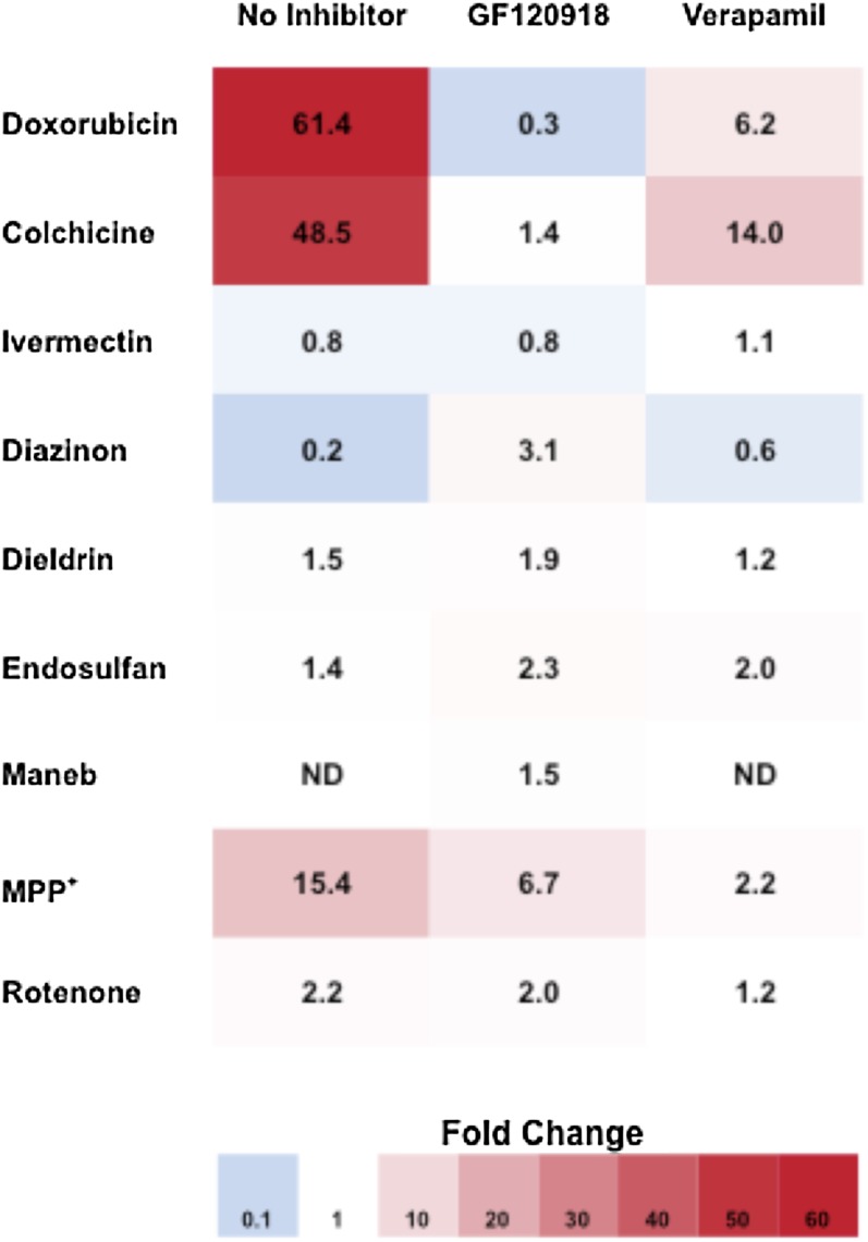
Fig. 4.
Visual representation of the fold change in cellular resistance to cytotoxic agents in LLC-MDR1-WT cells compared with LLC-vector cells. LLC-MDR1-WT cells display increased cellular resistance to P-gp substrates compared with LLC-vector cells, leading to fold changes greater than one (red). No difference between cellular sensitivities results in a fold change of one (white). A fold change of less than one (blue) indicates a result that is not P-gp mediated. Compounds were tested alone or in the presence of P-gp inhibitors (GF120918 and verapamil).

