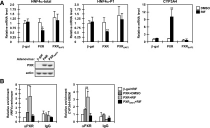
Fig. 4.

The AF2 domain determines the repression activity of PXR in the HNF4α P1 promoter. (A) HepG2 cells were infected with adeno-β-gal, adeno-hPXR, or adeno-hPXR∆AF2 for 30 hours and treated with DMSO or RIF for an additional 24 hours in FBS-free MEM. From those cells, total RNAs and whole-cell lysates were prepared and subjected to real-time PCR and Western blotting. The levels of total HNF4α, HNF4α P1 promoter-driven, and CYP3A4 mRNAs are expressed by taking the levels in the HepG2 cells infected with adeno-β-gal and treated with DMSO as one. Columns represent the mean ± S.D. from three independent experiments in triplicate. *P < 0.05 versus DMSO (Student’s t test); **P < 0.01 versus DMSO (Student’s t test). (B) After infection with the indicated adenovirus, HepG2 cells were treated with DMSO or RIF for 6 hours in FBS-free MEM and then subjected to ChIP assays with normal IgG or an anti-human PXR antibody as described in Materials and Methods. The relative enrichment of distal enhancer region of the HNF4a P1 promoter in the immunoprecipitated DNA fragments was determined by real-time PCR. The XREM of the CYP3A4 promoter was also analyzed as a gene that PXR upregulates. Values are normalized by amplification of sample inputs and expressed by taking the values in the HepG2 cells infected with adeno-β-gal and treated with DMSO as one. Columns represent the mean ± S.D. from three independent experiments. *P < 0.05 (Tukey-Kramer’s test); **P < 0.01 (Tukey-Kramer’s test).
