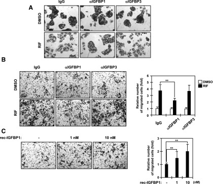
Fig. 7.

IGFBP1 is responsible for cell morphology and migration. (A) ShP51 cells were cotreated with normal IgG, an anti-IGFBP1 antibody, or an anti-IGFBP3 antibody and DMSO or RIF for 48 hours in FBS-free MEM and then stained with a crystal violet solution, as described in Materials and Methods. Scale bar, 100 μm. One representative out of three independent experiments is shown. (B and C) ShP51 cells were grown on the membrane of a transwell Boyden chamber and treated with the indicated stimuli for 48 hours in FBS-free MEM as described in Materials and Methods. Scale bar, 100 μm. The migrated cells were stained with a crystal violet solution and counted. Columns represent the mean ± S.D. from at least three independent experiments in triplicate. (B) Cells were cotreated with a normal IgG, an anti-IGFBP1 antibody, or an anti-IGFBP3 antibody and DMSO or RIF. **P < 0.01 (Dunnett’s test). (C) Cells were treated with PBS or a recombinant IGFBP1 protein at concentrations of 1 and 10 nM. **P < 0.01 (Dunnett’s test). rec, recombinant.
