Fig. 2.

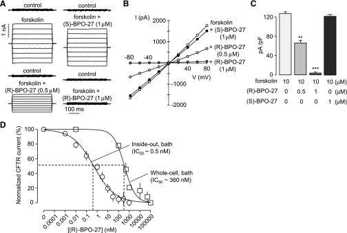
CFTR Cl− current measurements. (A) Whole-cell CFTR currents recorded in CHO-K1 cells at a holding potential of 0 mV and pulsing to voltages between ± 80 mV (in steps of 20 mV) in the absence and presence of (R)-BPO-27 (0.5 or 1 μM) or (S)-BPO-27 (1 µM). CFTR was stimulated by 10 μM forskolin. (B) Current/voltage (I/V) plot of mean currents at the middle of each voltage pulse. (C) Current density data measured at +80 mV (mean ± S.E., n = 5). (D) Whole-cell and macroscopic inside-out patch recordings were done in HEK-293T cells expressing wild-type CFTR. CFTR was stimulated by 10 μM forskolin in whole-cell recordings and by inclusion of 3 mM ATP and 10 U/ml protein kinase A catalytic subunit to the bath solution in inside-out patch recordings. Normalized CFTR current (mean ± S.E., n = 6–10) and IC50 values shown. **P < 0.01; ***P < 0.001.
