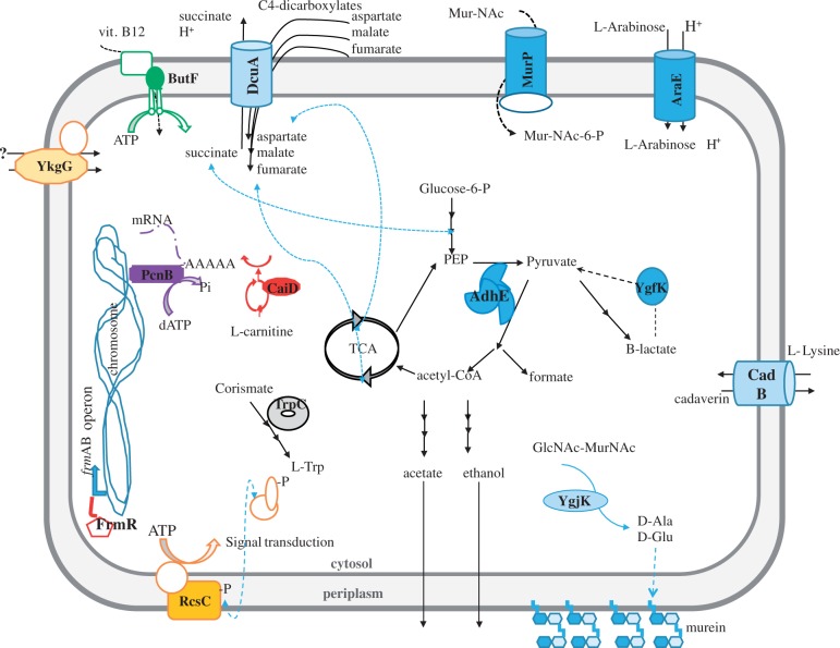
Fig. 6.
Subcellular localization of proteins mutated in the evolved populations under variable GroEL conditions. A cell-like structure with periplasmic space between inner- and outermembranes has been used to highlight the cellular position and affected pathways detected in evolving populations. The mutated proteins in the presence of groE overexpression are labeled in bold and indicated with solid color forms. When the affected protein belongs to a complex (e.g., the protein MurP), the other subunits are represented as empty forms with the same color lines. Colors are grouped by GO terms, so that all proteins belonging to the same molecular function are color-coded equally.

