Figure 7.
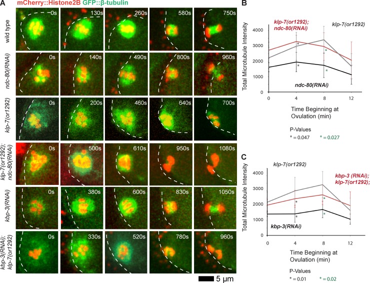
Disrupting the Ndc80 complex during meiosis I restores bipolarity to klp-7(−) oocytes but does not reduce microtubule accumulation. (A) Time-lapse spinning disk confocal images during meiosis I in live wild-type, ndc-80(−), kbp-3(−), and klp-7(−) single and double mutant oocytes expressing mCherry::Histone H2B and GFP::β-tubulin to mark chromosomes and microtubules, respectively. White dashed lines indicate the oocyte plasma membrane, and times after ovulation are indicated in time-lapse sequence frames. (B and C) Integrated GFP::β-tubulin pixel intensity values in arbitrary units beginning at ovulation during meiosis I (Materials and methods section Microscopy), comparing klp-7(or1292) (n = 10) with ndc-80(RNAi) (n = 3) and klp-7(or1292); ndc-80(RNAi) (n = 5; B) and kbp-3(RNAi) (n = 6) and kbp-3(RNAi);klp-7(or1292) (n = 6; C). All data are from video micrographs of individual oocytes, each isolated from different worms in multiple experiments.

