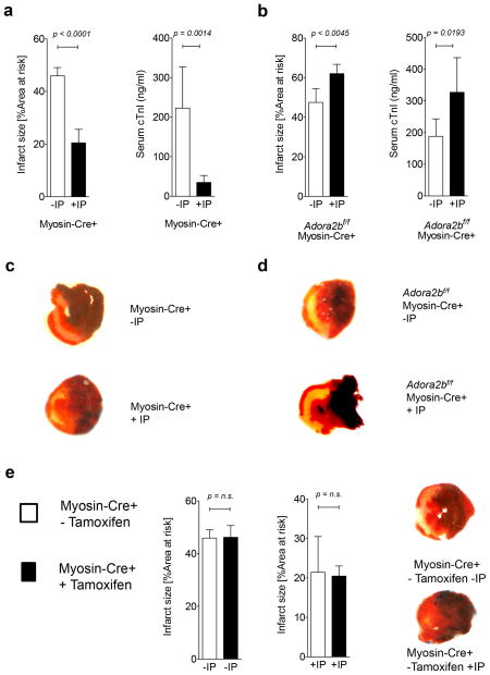
Figure 4. Effect of ischemic preconditioning on myocardial injury in cardiomyocyte-specific Adora2b-deficient mice.
(a–e) Mice underwent 60 min of ischemia with ischemic preconditioning (+IP; 4 cycles of 5 min of ischemia followed my 5 minutes of reperfusion) or without IP (−IP) followed by 120 minutes of reperfusion. Infarct sizes were measured by double staining with Evan’s blue and triphenyl-tetrazolium chloride. Infarct sizes are expressed as the percent of the area at risk (AAR) that underwent infarction. Serum troponin I concentrations were measured by enzyme-linked immunosorbent assay (ELISA). (a, b) Infarct sizes and serum troponin I levels in Myosin-Cre+ (controls) and Adora2bf/f-MyosinCre+ with and without IP. (c, d) Representative infarct staining from Myosin-Cre+ (controls) and Adora2bf/f-MyosinCre+. (e) Infarct sizes in Myosin-Cre+ (controls) with and without tamoxifen pretreatment. (n=3–6, ±SD).

