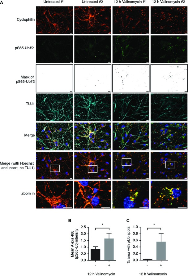
Figure 6.
pS65-Ub is induced in mouse primary neurons upon mitochondrial damage
Mouse primary neuronal cultures were left untreated or challenged with valinomycin as indicated.
- Two representative images are shown for each condition. Cells were fixed and stained with anti-cyclophilin F (mitochondria, red), anti-pS65-Ub#2 (green), anti-TUJ1 (cyan), and Hoechst (blue). Scale bars, 10 μm.
- Quantification of the mean intensity of the pS65-Ub signal in (A).
- The pS65-Ub mask shown in black/white in (A) is a binary image that was generated to quantify the area of the pS65-Ub spots.
Data information: Analyses were performed on five images for each condition for three independent experiments (two-sided unpaired t-test; *P < 0.05). Shown are mean values ± SD.

