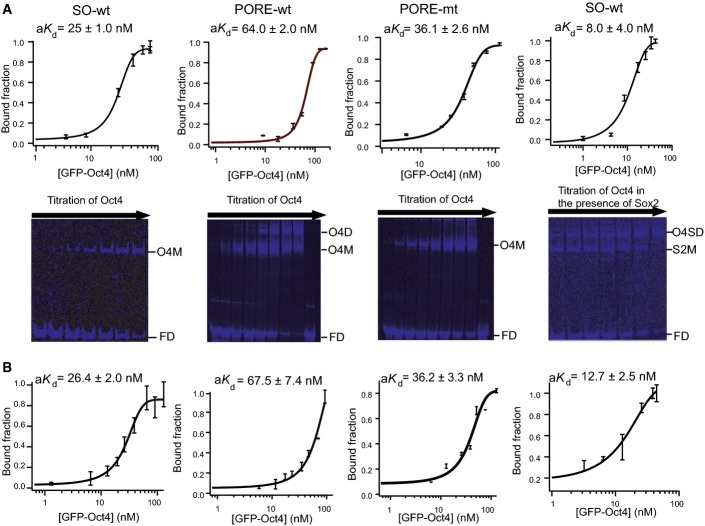
Figure 2.
Affinity of Oct4 binding to Sox/Oct and PORE motifs
- Titrations of GFP-Oct4 are shown from left to right with the Nanog Sox/Oct motif, the PORE-wt, the PORE-mt (a DNA probe in which one of the 2 binding sites in the PORE palindrome is mutated) and the Nanog Sox/Oct motif in the presence of mCherry-Sox2. Plots for quantitation of aKd are shown below (n = 3, mean ± SD). O4SD, GFP-Oct4 and Sox2 dimer complex; S2M, mCherry-Sox2 monomer complex; O4M, GFP-Oct4 monomer complex; FD, free DNA.
- Quantitation by FCS was performed independently (n = 3, mean ± SEM). All oligonucleotide sequences are in Appendix Table S1.
Source data are available online for this figure.

