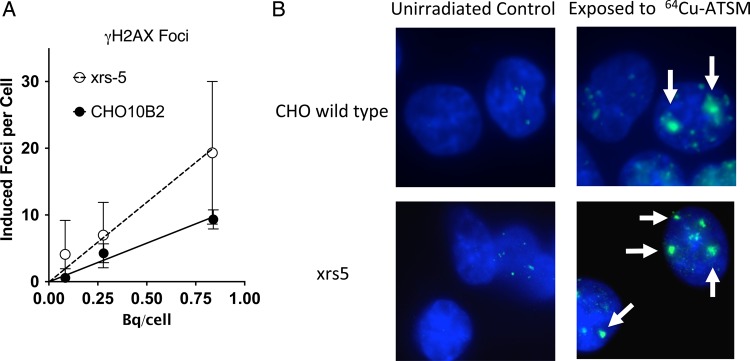
Fig. 2.

γH2AX foci formation after 64Cu-ATSM exposures. (A) Dose response of γH2AX foci formation after 64Cu-ATSM exposures. Foci response of cells incubated with varied activities normalized to background. Data points are the mean of at least three experiments. Error bars represent the standard error of the mean. (B) Examples of γH2AX foci formation in CHO wild-type and xrs5 cells without irradiation and after 0.84 and 0.83 Bq/cell of 64Cu-ATSM exposure. Arrows indicate cluster foci.
