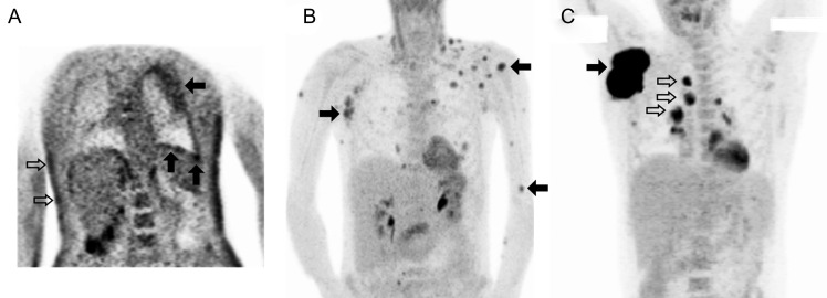
Figure 5.
Findings of 18F-fluorodeoxyglucose (18F-FDG) positron emission tomography (PET) in patients with intercurrent tumors, including primary effusion lymphoma (PEL), diffuse large B-cell lymphoma, and Kaposi sarcoma (KS). Representative 18F-FDG PET maximum intensity projections from 3 patients with KS herpesvirus–associated multicentric Castleman disease (KSHV-MCD) and intercurrent tumors, including large-cell lymphomas and KS. A, In a patient with KSHV-MCD and PEL of both pleural cavities and severe cutaneous KS, the pleural surface demonstrates moderately increased avidity throughout, with patches of intense uptake (closed arrows; pleural nodal maximal standardized uptake value [SUVmax], 7.0), whereas the lymphomatous effusion (above lower closed arrows) is not 18F-FDG avid. Areas of moderate hypermetabolic activity are also seen in the skin at sites of KS involvement (open arrows). Lymph nodes and spleen were metabolically normal. The acquired imaging field and image quality in this patient was reduced owing to his critical illness. B, In a patient with KSHV-MCD and extracavitary PEL presenting with cutaneous and soft tissue, marked areas of avidity are seen at the sites of extracavitary PEL (closed arrows; lesion SUVmax, 11.0), while nodes are normal; the patient had previously undergone a splenectomy. C, In a patient with KSHV-MCD and diffuse large B-cell lymphoma, intense avidity is seen at the primary lymphomatous mass (closed arrow; SUVmax, 38.2) and less intense avidity at nearby involved nodes (open arrows), whereas uninvolved nodes and spleen are normal. Normal cardiac uptake is seen in B and C, and normal tracer excretion into the renal collecting system in B.

