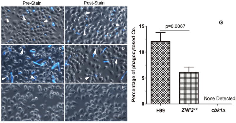
Figure 2.

Phagocytosis of cryptococcal cells of different morphology by murine macrophage cells. Murine J774A.1 cells were infected by C. neoformans wild-type strain H99 (A, B), the PCTR4-2ZNF2 strain (C, D), and the cbk1Δ mutant (E, F) for two hours and then fixed. The left panel images were taken with cryptococcal cells stained with calcofluor white prior to the infection (A, C, E). The right panel images were taken with the co-culture stained with calcofluor white after phagocytosis (B, D, F). Statistical analysis was performed on the percentage of phagocytosed C. neoformans (Cn) cells (G). A p value lower than .05 is considered statistically significant. No cbk1Δ mutant cells were phagocytosed and thus no fluorescent signal was detected.
