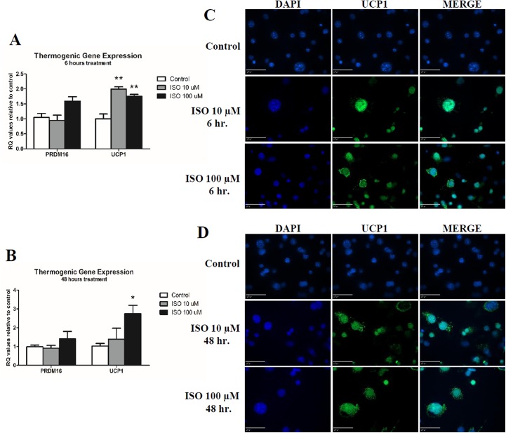
Fig 2. Quantitative PCR Analysis and Immunofluoresence of Thermogenic Markers in Mature Adipocytes.
Mature 3T3-L1s were treated with either 10 or 100 μM isoproterenol for 6 and 48 hours before isolation of mRNA for qPCR (A & B respectively). Three biological replicates and technical replicates were used and data were normalized to β-Actin and control (no treatment). Statistics were performed using t-tests. Statistical differences between control and treatment, unless otherwise designated, are indicated with * p<0.05 and ** p<0.01. For immunoflouresence, cells were co-stained with anti-UCP1 in green following 6 or 48 hours of isoproterenol treatment (C & D respectively). Nuclear staining was performed using DAPI shown in blue. Scale bar equals 50 μM distance.

