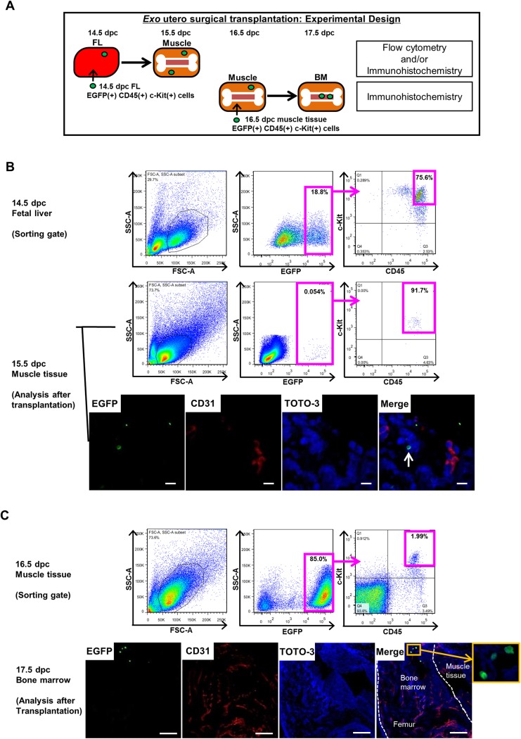
Fig 5. Fetal CD45(+) c-Kit(+) cells migrate from liver to muscle and then to BM.
Fetal CD45(+) c-Kit(+) cell migration was assessed by exo utero surgical transplantation. (A) Experimental design showing the transplantation protocol. EGFP(+) CD45(+) c-Kit(+) cells were sorted from FL of EGFP Tg mouse embryos at 14.5 dpc and from muscle tissues surrounding femurs at 16.5 dpc and transplanted into corresponding tissues of recipient C57BL/6 mouse embryos at the same developmental stage. After 24 hours, the presence of EGFP(+) cells in muscle tissue and BM was analyzed by flow cytometry and/or immunohistochemistry. (B) Flow cytometric profile exhibiting gate setting used to sort EGFP(+) CD45(+) c-Kit(+) cells from FL of EGFP Tg embryos at 14.5 dpc (upper). At 24 hours after transplantation, muscle tissue was dissociated into single cells and analyzed by flow cytometry. Representative flow cytometric profile shows EGFP(+) CD45(+) c-Kit(+) cells present in muscle tissue of 15.5 dpc recipients (middle). Muscle tissue was sectioned and immunostained with CD31 (red) and TOTO-3 iodide (blue). Representative confocal image showing EGFP(+) donor cells (arrow; green) in muscle (lower). Scale bar represents 10 μm for all panels. (C) Flow cytometric profile exhibiting gate setting used to sort EGFP(+) CD45(+) c-Kit(+) cells from muscle tissue of EGFP Tg embryos at 16.5 dpc (upper). At 24 hours after transplantation, femurs were sectioned and immunostained with CD31 (red) and TOTO-3 iodide (blue). Scale bar represents 100 μm. EGFP(+) donor cells (green) are present in BM at 17.5 dpc. Boxed area of merge panel is shown at higher magnification.

