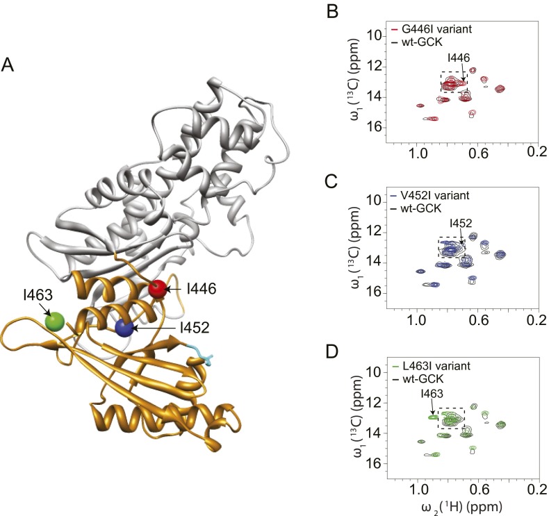
Fig. S3.
Positional insertion of 13C-Ile to probe the structure of the small domain α13-helix of unliganded GCK. (A) Location of Ile insertions; (B–D) 1H-13C HMQC spectra of the G446I, V452I, L463I variants of GCK demonstrate the appearance of new cross-peaks. The boxed area denotes the region expected for disordered isoleucines.

