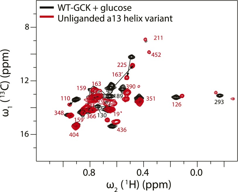
Fig. S5.
Overlay of 1H-13C Ile HMQC spectra of glucose-bound wild-type GCK with the unliganded α13-helix variant. In the unliganded α13-helix variant, I159 and I163 each display two peaks. One is in the disordered region corresponding to their position in the glucose bound wild-type, whereas the other peak is shifted away from the disordered region (denoted with a prime).

