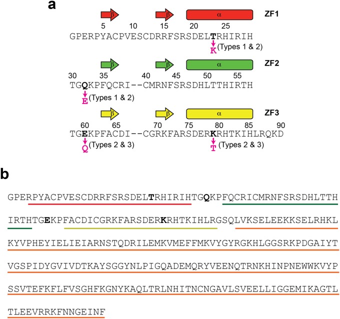
Fig S7.
The Egr-1 ZF protein constructs used in this study. (A) The amino acid sequence of the Egr-1 ZF-DBD constructs. Each construct is a 91-residue protein containing 3 ZFs. The first two residues are from the expression vector. The residue numbering is according to Pavletich and Pabo (37). The sequences of individual ZFs are aligned, and the mutation sites for type 1, 2, and 3 mutant proteins are indicated. (B) The amino acid sequence of the type 0 Egr-1 ZFN. Regions of Egr-1 ZF1, ZF2, ZF3, and FokI ND are indicated by red, green, yellow, and orange lines, respectively. The mutation sites for type 1, 2, and 3 constructs are shown in bold.

