Fig. 4.
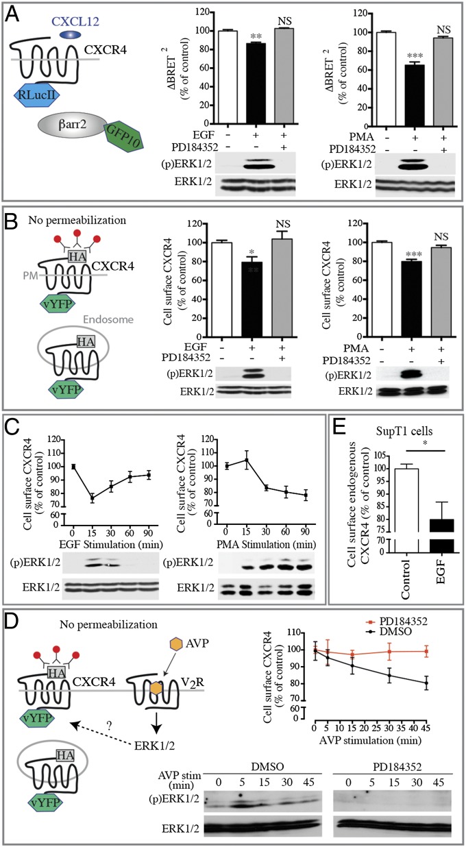
Acute pharmacological activation of the ERK/MAPK pathway blunts CXCR4 signaling by reducing receptor cell-surface localization. (A) CXCL12-promoted βarr2 translocation is measured by BRET in HEK293T cells transfected with CXCR4-RLucII and βarr2-GFP10. Cells were pretreated with PD184352 for 30 min before stimulation with EGF for 15 min or with PMA for 30 min. BRET400-GFP10 between CXCR4-RLucII and βarr2-GFP10 was measured after the addition of coel-400a, 15 min following the addition of CXCL12. (B) CXCR4 cell-surface expression levels were assessed by dual-flow cytometry in HEK293T cells stably expressing HA-CXCR4-vYFP that were pretreated or not with PD184352 for 30 min before stimulation with EGF for 15 min or with PMA for 30 min. (C) Kinetics of CXCR4 cell-surface level reduction and ERK1/2 activation following EGF or PMA treatments. (D) Kinetics of the reduction of CXCR4 cell-surface levels and ERK1/2 activation in HEK293T cells transfected with HA-CXCR4-vYFP and myc-V2R pretreated or not with PD184352 for 30 min before stimulation with AVP for the indicated time. (E) CXCR4 cell-surface expression level in SupT1 cells treated or not with EGF for 15 min. In all cases, data shown represent the mean ± SEM of at least three independent experiments and are normalized to 100% of the control conditions. Total ERK1/2 and ERK1/2 activation status [(p)ERK1/2] were assessed by immunoblotting. *P < 0.05; **P < 0.01; ***P < 0.001; NS, not significant.

