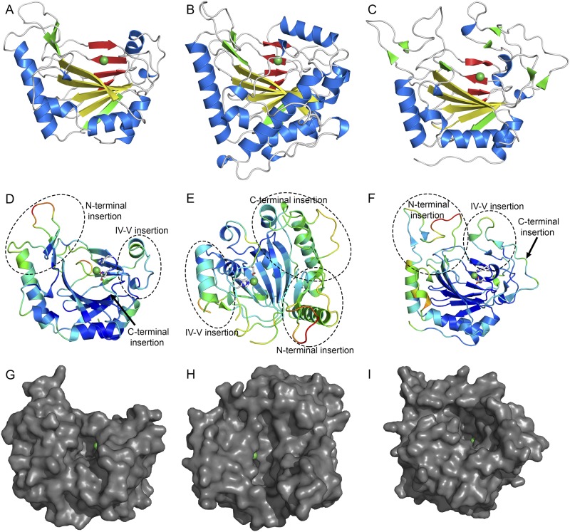
Fig. S2.
Orthosomycin-associated oxygenase structures. Cartoon representations of AviO1 (A), EvdO1 (B), and EvdO2 (C) in the same orientations. The major sheet is colored yellow, the minor sheet is red, and nickel ions are shown as green spheres. (D–F) Cartoon representation of AviO1 (D), EvdO1 (E), and EvdO2 (F) colored by crystallographic temperature factors. Warm colors indicate high B-factors and more thermal motion. Three insertion sites common to the PhyH subfamily of AKG/Fe(II)-dependent oxidases are labeled. The orientation of each maximizes the view of the three insertions and the binding site. (G–I) Surface representation of AviO1 (G), EvdO1 (H), and EvdO2 (I) showing the prime substrate binding site in the same orientation as used in D–F.

