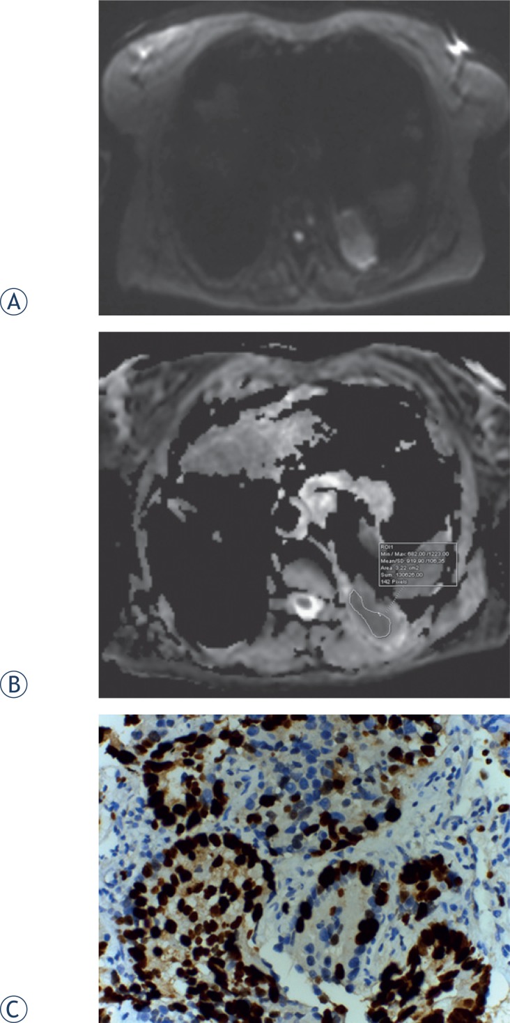
FIGURE 1.
Diffusion-weighted (DW)-MRI, apparent diffusion coefficient (ADC) map of a 62-year-old female with adenocarcinoma. (A) Tumour shows heterogeneously high signal intensity on DW-MRI, for which the b value is 800 s/mm2. (B) On the ADC map, the tumour demonstrates heterogeneous diffusion restriction. (C) Proli ferative index 95% in glandular epithelium (Ki-67X400).

