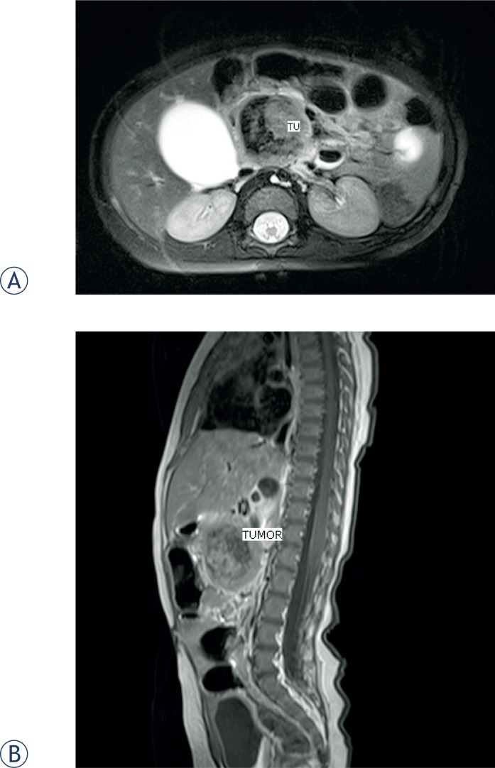
FIGURE 2.
MRI confirmed well circumscribed tumor mass, with a diameter of 37 mm. The origin of the mass was in the head of the pancreas and in the uncinate process. There was no infiltration of the surrounding tissue. Tumor impressed the caval vein and pushed the superior mesenteric artery and vein ventraly and lateraly. (A) Coronal plane. (B) Sagital plane.

