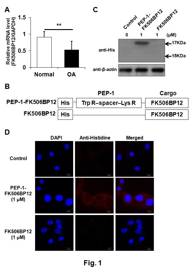
Fig. 1. Transduction of PEP-1-FK506BP12 into human chondrocytes. (A) Expression of FK506BP12 in normal and OA cartilages. GAPDH served as an internal control. Data are expressed as the means ± SDs of data from triplicate experiments using cartilage from three different donors. **P < 0.01 vs. normal cartilage. (B) Schematic structure of PEP-1- FK506BP12 and FK506BP12. PEP-1-PTD, a 21-residue peptide carrier, consists of hydrophobic Trp R (tryptophan-rich), spacer, and hydrophilic Lys R (lysine-rich) domains. His, the 6-histidine motif used in the purification and detection of the two proteins. (C) Transduction of PEP-1-FK506BP12 into human primary chondrocytes. Chondrocytes were incubated with PEP-1-FK506BP12 or FK506BP12 (1 μM) for 2 h and then with trypsin-EDTA for 1 h. PEP-1-FK506BP12 and FK506BP12 levels in the cell lysates were measured by Western blotting. Control, untreated chondrocytes. (D) Distribution of transduced PEP-1-FK506BP12 in human primary chondrocytes. Chondrocytes were incubated for 2 h with PEP-1-FK506BP12 or FK506BP12 (1 μM). Control, untreated cells. Nuclei were stained with DAPI. Magnification, ×600.

