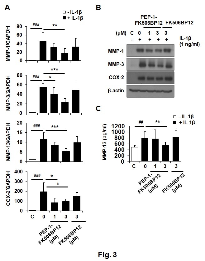
Fig. 3. PEP-1-FK506BP12 suppresses IL-1β-induced MMP expression in chondrocytes. (A) Relative expression levels of MMP-1, -3, and -13; and COX-2 mRNA, in chondrocytes treated with PEP-1-FK506BP12 or FK506BP12. Chondrocytes were pretreated with PEP-1-FK506BP12 (1 and 3 μM) or FK506BP12 (3 μM) for 2 h and exposed to IL-1β (1 ng/ml) for 6 h. mRNA levels were measured via rt-qPCR. (B), (C) Relative expression levels of MMP-1, -3, and -13; and COX-2 proteins, in chondrocytes treated with PEP-1-FK506BP12 (1 and 3 μM) or FK506BP12 (3 μM). Chondrocytes were incubated with PEP-1-FK506BP12 or FK506BP12 for 2 h and then stimulated with IL-1β (1 ng/ml) for 24 h. (B) MMP-1, and -3; and COX-2 protein levels, were measured by Western blotting. A representative blot from experiments performed using cartilage from three different donors is shown. (C) Relative levels of MMP-13 released into culture media. Data are the means ± SDs of data from duplicate experiments using cartilage from three different donors. ##P < 0.01, ###P < 0.005 vs. IL-1β-untreated chondrocytes. *P < 0.05, **P < 0.01, ***P < 0.005 vs. IL-1β-treated chondrocytes.

