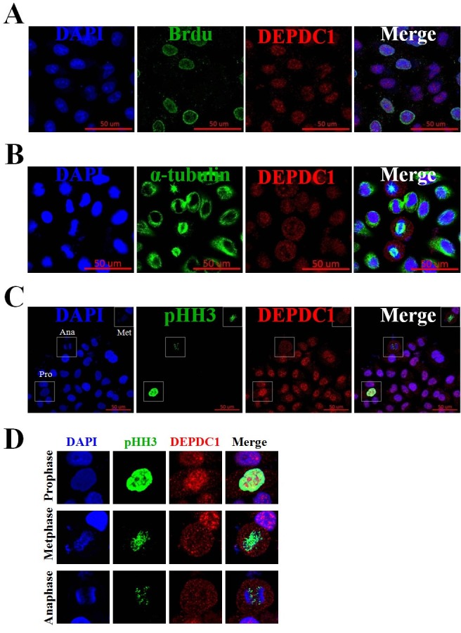
Fig. 2. Immunofluorescence assays of DEPDC1 expression during cell cycle. (A) PRR11 expression in S phase of the cell cycle. Asynchronously growing HeLa cells were incubated in the presence of BrdU for 30 min and then stained simultaneously with anti-BrdU and anti-DEPDC1 antibodies. Cell nuclei were stained with DAPI (blue). Scale bar, 50 μm. (B) and (C) Expression of DEPDC1 in M phase of the cell cycle. Exponentially growing cells were fixed and simultaneously stained with anti-DEPDC1 (red) and anti-α-tubulin (green) or anti-phospho-histone H3 (green). Cell nuclei were stained with DAPI (blue). (D) Higher magnification images of DEPDC1 expression in prophase, metaphase and anaphase as indicated in (C).