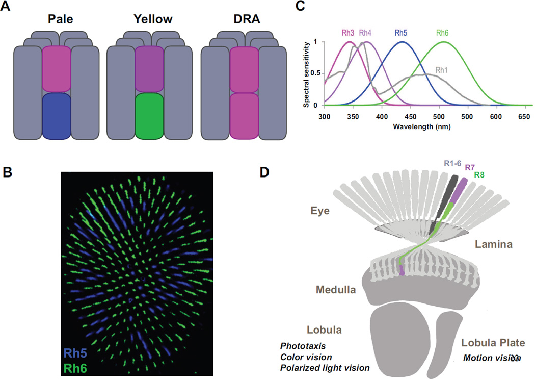
Figure 1. The eye and the optic lobe of adult Drosophila.
A A single ommatidium contains eight photoreceptors, six outers R1-6 (grey) and two inners R7-8 (colors). Outers express the Rh1 opsin, R7s express either Rh3 or Rh4 while R8s express Rh5 or Rh6. Four types of ommatida are found in the eye. ‘Pale’ and ‘yellow’ ommatidia are distributed stochastically in the main part of the eye. In the dorsal third, pale and specialized dorsal third yellow subtypes are found (not shown). In one to two rows of ommatidia in the dorsal rim are (DRA) of the retina, the remaining DRA subtypes are found. B Stochastic distribution of Rh5 (blue) and Rh6 (green) expressing R8s in the main part of the eye. C Normalized spectral preference curves of the different rhodopsins expressed in the eye of the fly. Rh1 shows broad spectral sensitivity peaking in both the blue and the UV due to the presence of a sensitizing pigment (Modified from 3) D Photoreceptors project to the optic lobe. Outer photoreceptors send their axons to the lamina while R7/R8 photoreceptors send theirs to the medulla. The lobula is involved in spectral preference, color and polarized light vision. The lobula plate is a center for motion detection.

