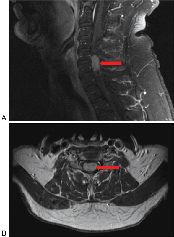
Fig. 2.

(A) Sagittal T1-weighted post–gadolinium contrast cervical magnetic resonance image illustrating the presence of a homogeneously enhancing ependymoma at the levels of C5 and C6 (arrow). (B) Corresponding axial view noting the centromedullary location of the ependymoma (arrow).
