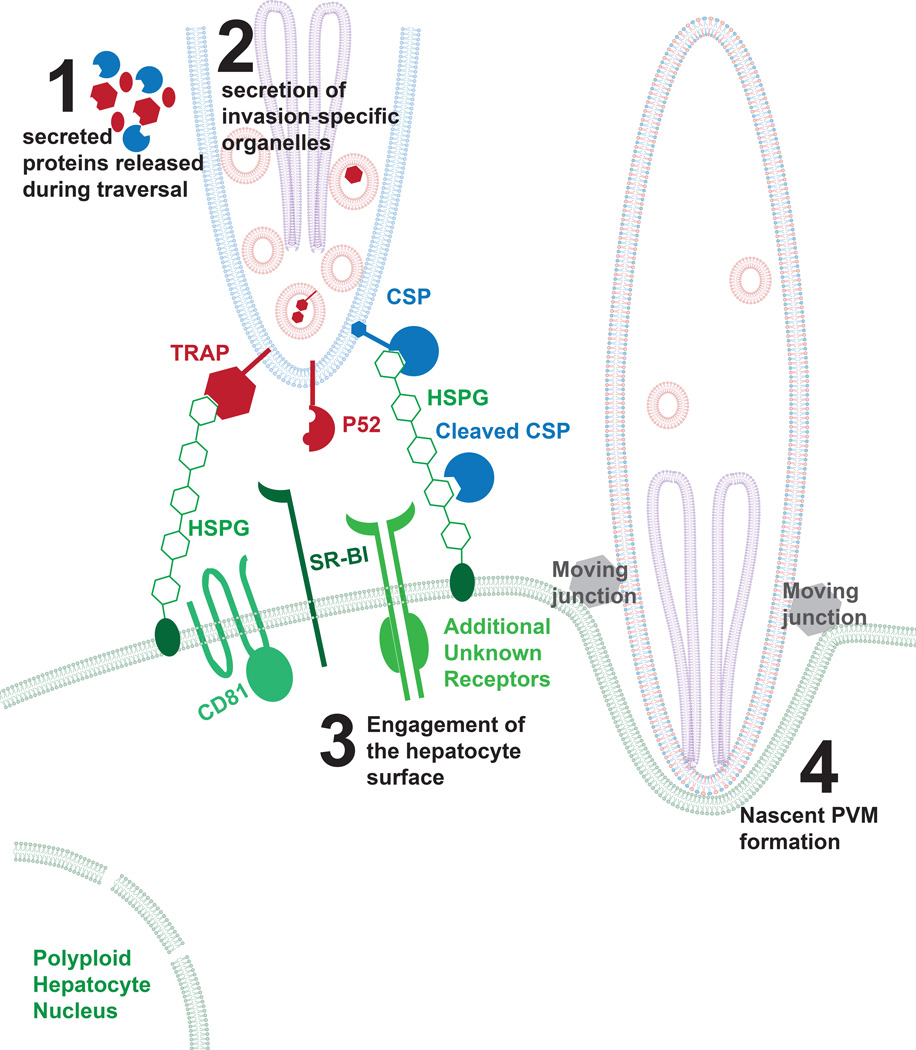
Figure 1. Model of initial attachment and invasion of the Plasmodium sporozoite.
After transmission, sporozoites glide and traverse through the skin into the blood stream, which involves the secretion of micronemal proteins. After sporozoites traverse the sinusoids, they directly engage a hepatocyte for invasion. This binding likely involves several factors including the interaction between CSP and highly sulfated proteoglycans. For most species of Plasmodium, this requires the expression of CD81 and SR-BI, although there has been no evidence of direct interaction between parasite proteins and these molecules. After attachment, sporozoites initiate the moving junction and the formation of the nascent parasitophorous vacuole membrane. The sporozoite plasma membrane is shown in blue, the micronemes in red, the rhoptries in purple and hepatocyte membranes in green. A red and blue co-colored membrane (shown in step 4) is indicative of the sporozoite membrane after it has been modified by micronemal proteins.

