Figure 8.
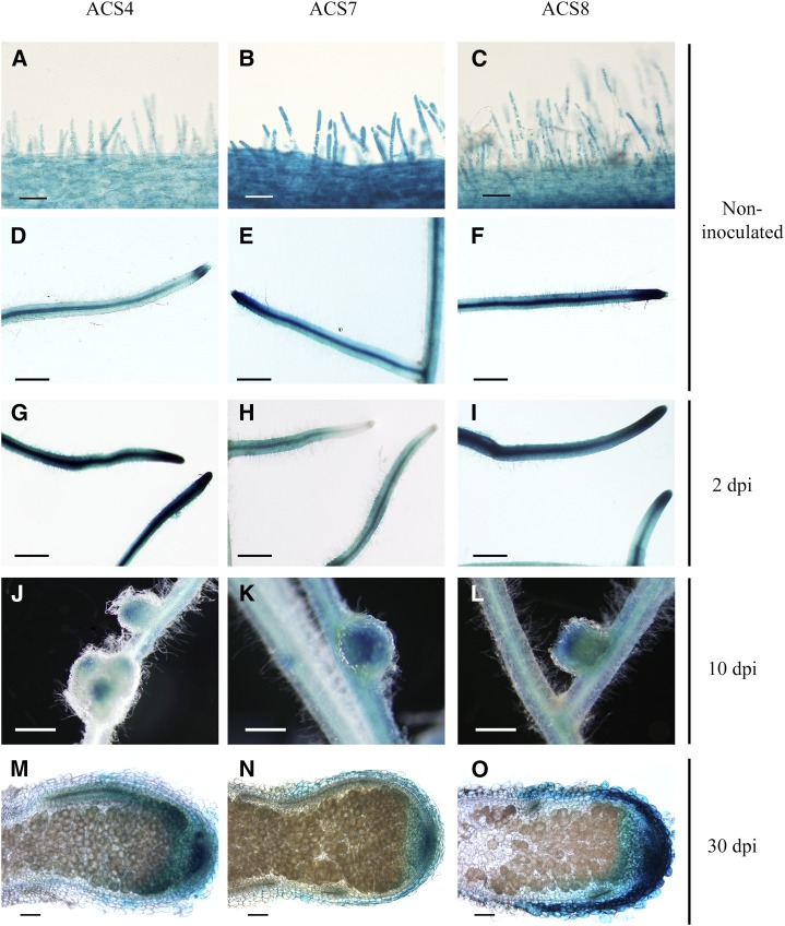
Histochemical localization of the expression of the three symbiosis-responsive ACS genes in M. truncatula roots at different stages after inoculation. The promoter of three ACS genes was cloned in fusion with the GUS reporter gene in the pLP100 binary vector. M. truncatula roots were transformed by hairy root transformation. The expression of gene ACS4 is represented in A to M, ACS7 in B to N, and ACS8 in C to O. A to F, Bright-field images of 5-bromo-4-chloro-3-indolyl-β-glucuronic acid (X-Gluc)-stained roots before inoculation. G to I, Roots at 24 hpi. J to L, Roots at 10 dpi. M to O, Roots at 30 dpi. Bars = 25 µm (A–C), 500 µm (D–L), and 100 µm (M–O).

