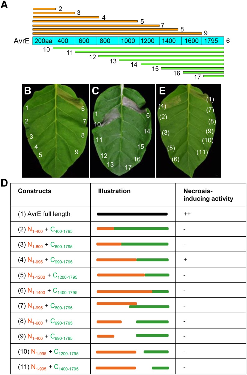
Figure 3.
Serial deletion analysis reveals a putative two-domain structure of AvrE. A, Diagram showing the AvrE deletion constructs used in this study. B and C, Necrosis-inducing activity of AvrE proteins transiently expressed in tobacco leaves. Numbering is as follows: 1, empty vector; 2, AvrE1-200aa; 3, AvrE1-400aa; 4, AvrE1-596aa; 5, AvrE1-995aa; 6, His:AvrE; 7, AvrE1-1200aa; 8, AvrE1-1400aa; 9, AvrE1-1600aa; 10, AvrE200-1795aa; 11, AvrE400-1795aa; 12, AvrE600-1795aa; 13, AvrE800-1795aa; 14, AvrE990-1795aa; 15, AvrE1200-1795aa; 16, AvrE1400-1795aa; and 17, AvrE1600-1795aa. Necrosis is characterized by tissue collapse and discoloration (e.g. gray color) in the infiltrated area. D, Combinations of AvrE deletion derivatives used for transient coexpression experiments in tobacco. For each combination, two Agrobacterium tumefaciens strains (optical density at 600 nm [OD600] = 0.1) were mixed at a 1:1 ratio and infiltrated into tobacco leaves. E, Necrosis (indicated by tissue collapse with gray color) induced by combinations of truncated AvrE proteins. Photographs were taken 2 d after 10 µm DEX spray. Numbering is the same as shown in D.

