Figure 2.
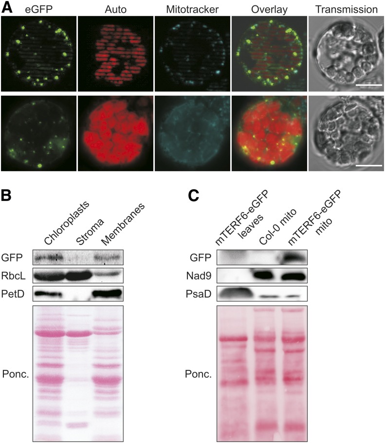
The mTERF6 protein is targeted to chloroplasts and mitochondria. A, The mterf6-1 mutant was stably transformed to express an mTERF6-eGFP fusion protein, and expression of the fusion was monitored in protoplasts of positive transformants using fluorescence microscopy. The eGFP signal reveals the distribution of the fusion protein, autofluorescence of chlorophyll (red) pinpoints chloroplasts (Auto), and Mitotracker signals (cyan) visualize mitochondria (Mitotracker). Bars = 10 µm. B, The mTERF6 protein is targeted to chloroplasts. Chloroplast, stroma, and membrane fractions were isolated from complemented mterf6-1 (mTERF6-eGFP) plants, subjected to SDS-PAGE, transferred to a polyvinylidene difluoride membrane, and exposed to antibodies raised against GFP (to detect the mTERF6-GFP fusion protein), RbcL (as a control for stromal proteins), or PetD (as a control for the membrane fraction). C, The mTERF6 protein is targeted to mitochondria. A whole-leaf extract from complemented 4-week-old mterf6-1 (mTERF6-eGFP) plants and mitochondria-enriched fractions (mito) from Col-0 and mTERF6-eGFP plants were subjected to SDS-PAGE, transferred to a polyvinylidene difluoride membrane, and exposed to antibodies raised against GFP (to detect the mTERF6-GFP fusion protein), Nad9 (as a control for mitochondrial proteins), or PsaD (as a control for chloroplast proteins). Ponc., Ponceau Red.

