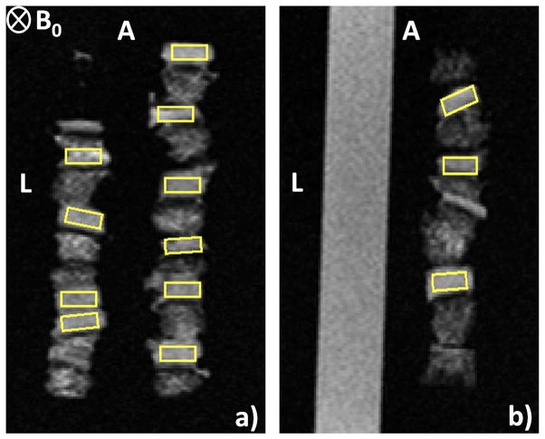
Figure 2.
T2 W (2D spin-echo) of TE = 20 ms images of cartilage samples with ROIs (yellow color). The 4-well ULTEM sample holder contained six plugs per well in three wells, and DPBS in the fourth well. The ‘anterior’ and ‘left’ sample positions are indicated by the letters ‘A’ and ‘L’, respectively, while the static magnetic field, B0, is oriented into the image plane. (a) Slice 1, intersecting two of the wells containing cartilage. (b) slice 2, intersecting the third cartilage-containing well and the DPBS-containing well.

