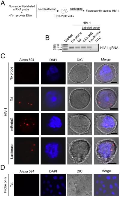
Fig. 2.
Labeling of HIV-1 particles via packaging of fluorescent mRNAs and the pattern within infected cells. (A) Flow diagram showing the steps for preparing labeled HIV-1 by packaging of fluorescent mRNA probes. Probes were prepared by in vitro transcription using plasmid cDNAs and the resulting mRNAs were labeled by covalent conjugation with the Alexa Fluor® 594 red-fluorescent dye. Fluorescently-labeled mRNA probes and a proviral DNA plasmid encoding the HIV-1 NL4-3 clone were co-transfected into HEK-293T cells, resulting in the packaging of labeled mRNAs into progeny virions during HIV-1 particle assembly. (B) RT-PCR detection of HIV-1 genomic RNA (gRNA) in the supernatants of cells transfected as described in A. HIV-1 particles were generated with or without labeled mRNA probes (no probe). Three labeled mRNA probes of different origins were made and packaged into virus: full-length HIV-1 Tat1-86 mRNA, and mRNAs from the C-terminal portions of mouse EndoG (mEndoG) and firefly luciferase. For an RT-PCR reaction control, reverse transcription of DNase-treated RNA without the addition of reverse transcriptase was input to serve as a negative control (NTC, no cDNA template control). Marker indicates 100 base pair (bp) DNA ladder marker where the 100, 200 and 300 bp markers are shown. (C) Confocal images of Z-stack sections showing a distinct fluorescent pattern of punctate, focal signals within HeLa MAGI-CCR5 cells infected by the indicated probe-labeled (red) or unlabeled HIV-1 particles. Representative cells are shown. DAPI (blue) staining indicates cell nuclei. DIC (differential interference contrast) images show cell shape. Note that the size of the fluorescent spots does not correspond to the diameter of a viral particle but reflects scattering of photons in the detector. Scale bar = 5 μm. (D) Representative field in which labeled Tat mRNA alone was added to HeLa MAGI-CCR5 cells for 24 h in culture imaged at 63× magnification. (For interpretation of the references to color in this figure legend, the reader is referred to the web version of this article.)

