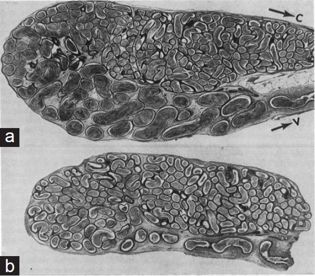
Figure 5.

(a) Histological section of the scrotal cauda epididymidis of a rat. (b) A comparable section of the ipsilateral cauda reflected the abdomen 3 months previously. In (b) the diameter and length of the distal segment are reduced. Note that the epididymis in (b) remained in continuity with a normal scrotal testis, and had a normal sperm number in the caput. C: The lower region of the corpus epididymidis. V: the vas deferens.
