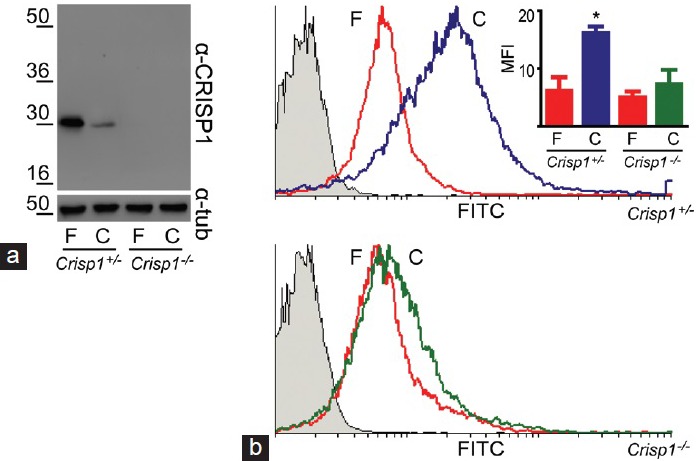
Figure 1.

(a) Fate of CRISP1 during mouse sperm capacitation. Total protein extracts from equal amounts of fresh (F) and capacitated (C) epididymal spermatozoa collected from either Crisp1+/− or Crisp1−/− animals were analyzed by SDS-PAGE and Western blots using an anti-CRISP1 antibody (α-CRISP1). β-tubulin was used as control of loading (α-tub). (b) Evaluation of tyrosine phosphorylation in CRISP1-null spermatozoa. Fresh (F) and capacitated (C) epididymal spermatozoa from Crisp1+/− or Crisp1−/− animals were subjected to flow cytometric analysis with an anti-phospho tyrosine antibody. The figure shows representative histograms for each genotype. The gray histograms correspond to sperm cells incubated with normal IgG as primary antibody (negative control). The mean fluorescence intensity (MFI) ± s.e.m. of three experiments is shown as an inset, *P < 0.05.
