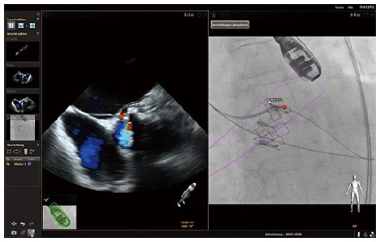
Figure 6.

A case of a prosthetic paravalvular leak presenting aortic regurgitation after transcatheter aortic valve repair. The left panel demonstrates the Echo image in a short axis view, presenting the aortic annulus and depicting the regurgitant jet with Color Doppler. Here the marker was placed incorrectly, consequently transfering the marker next to the destination in the fluoroscopic image in the right panel. The red marker demonstrates where the region of interest should have been defined, reflecting the correct position in the fluoroscopic image.
