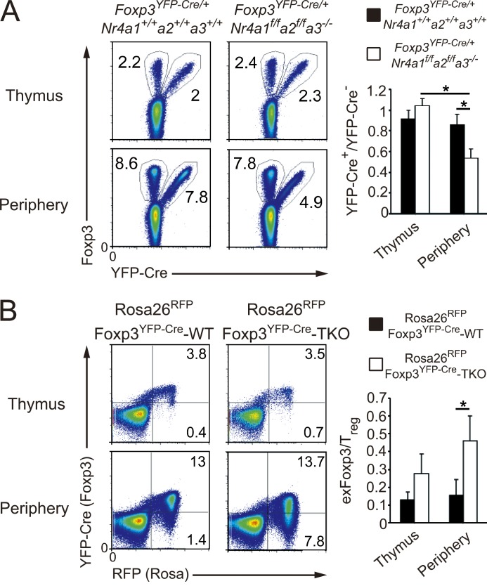
Figure 7.
Accelerated loss of Foxp3 expression and conversion to exFoxp3 cells of Nr4a-TKO T reg cells. (A, left) Flow cytometry of CD4-SP thymocytes and CD4+ T cells from pooled spleens and lymph nodes (Periphery) in 4-wk-old Foxp3YFP-Cre/+Nr4a1+/+Nr4a2+/+Nr4a3+/+ and Foxp3YFP-Cre/+Nr4a1f/fNr4a2f/fNr4a3−/− mice. (right) Quantification of the flow cytometry results. Ratios of YFP+/YFP− in total Foxp3+ cells are shown (n = 4 mice per genotype, pooled from 3 independent cohorts per genotype, mean and SD). (B, left) Flow cytometry of CD4-SP thymocytes and peripheral CD4+ T cells from 4-wk-old Rosa26RFPFoxp3YFP-Cre-WT and Rosa26RFPFoxp3YFP-Cre-Nr4a-TKO mice. (right) Quantification of the flow cytometry results. The ratios of exFoxp3/T reg cells within CD4+RFP+ cells from the thymus and periphery of 4–5-wk-old mice were quantified (n = 4 mice per genotype, pooled from 3 independent cohorts per genotype, mean and SD). *, P < 0.05 (one-way ANOVA with Bonferroni test).

