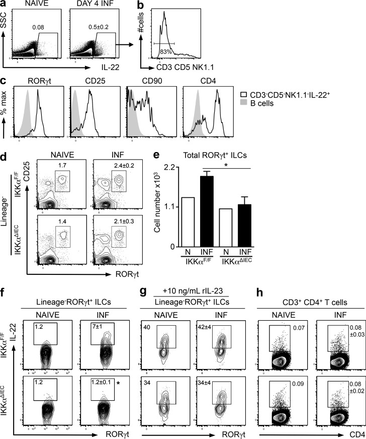
Figure 4.
C. rodentium infection–induced ILC-dependent IL-22 responses are diminished in IKKαΔIEC mice. IKKαF/F mice were infected (INF) with C. rodentium and sacrificed at day 4 p.i. (a) Representative plots displaying frequencies of IL-22+ cells in the mLNs. (b) Expression of T cell (CD3 and CD5) and NK cell (NK1.1) markers among IL-22+ cells. (c) Expression of ILC surface markers in gated CD3−CD5−NK1.1−IL-22+ cells (black, open histograms) compared with CD19+ B cells (gray, closed histograms). (d) Representative plots displaying frequencies of CD3−CD5−CD19−CD11c−NK1.1−, RORγt+CD25+ ILCs in the mLNs of naive and C. rodentium–infected littermate control IKKαF/F and IKKαΔIEC mice. (e) Total mLN ILC3s. (f) Frequencies of IL-22–expressing RORγt+ ILCs in the mLN after ex vivo PMA and ionomycin stimulation. (g) Frequencies of IL-22–expressing RORγt+ ILCs in the mLNs after stimulation with rIL-23, PMA, and ionomycin. (h) Frequencies of CD3+CD4+ T cells in the mLNs expressing IL-22 after ex vivo PMA and ionomycin stimulation. Data for a–f and h are representative of five experiments (IKKαF/F, total n = 21; IKKαΔIEC, n = 17 + 1 naive mouse of each genotype per experiment), and data for g are representative of three experiments (IKKαF/F, total n = 13; IKKαΔIEC, n = 11 + 1 naive mouse of each genotype per experiment). Data are expressed as mean ± SEM. *, P < 0.05 compared with IKKαF/F INF.

