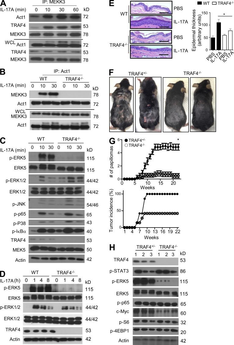
Figure 4.
TRAF4 is a bridge protein for the Act1-mediated MEKK3–ERK5 pathway. (A) WT kidney epithelial cells were treated with IL-17A for the indicated times. Lysates were then immunopreciptated with anti-MEKK3 antibody, followed by Western blot analysis. (B) TRAF4 WT and TRAF4−/− kidney epithelial cells were treated with IL-17A for the indicated times. Cell lysates were then immunopreciptated with anti-Act1 antibody, followed by Western blot analysis. (C) TRAF4 WT and TRAF4−/− primary keratinocytes were treated with IL-17A, followed by Western blot analysis. (D) Western blot analysis of epidermal lysates from ears of WT and TRAF4−/− mice injected with IL-17A for the indicated times. (E) H&E staining of ear sections from TRAF4 WT or TRAF4−/− mice injected intradermally with IL-17A or PBS alone. Graph represents mean epidermal thickness (arbitrary units) ± SEM. *, P < 0.05 (five fields were analyzed, Student’s t test). Bars, 50 µm. (F) Photographs of TRAF4+/− and TRAF4 −/− mice treated with DMBA/TPA for 22 wk. (G) Tumor numbers and tumor incidence of DMBA/TPA-treated TRAF4+/− and TRAF4−/− mice (n = 7 mice per group). Tumor numbers presented are the mean number of tumors per mice ± SEM at different time points. *, P < 0.05 (two-way ANOVA). (H) Western blot analysis of the tumor tissue from TRAF4+/− and TRAF4−/− mice. Each lane represents an independent sample. All experimental data were verified in at least three independent experiments.

