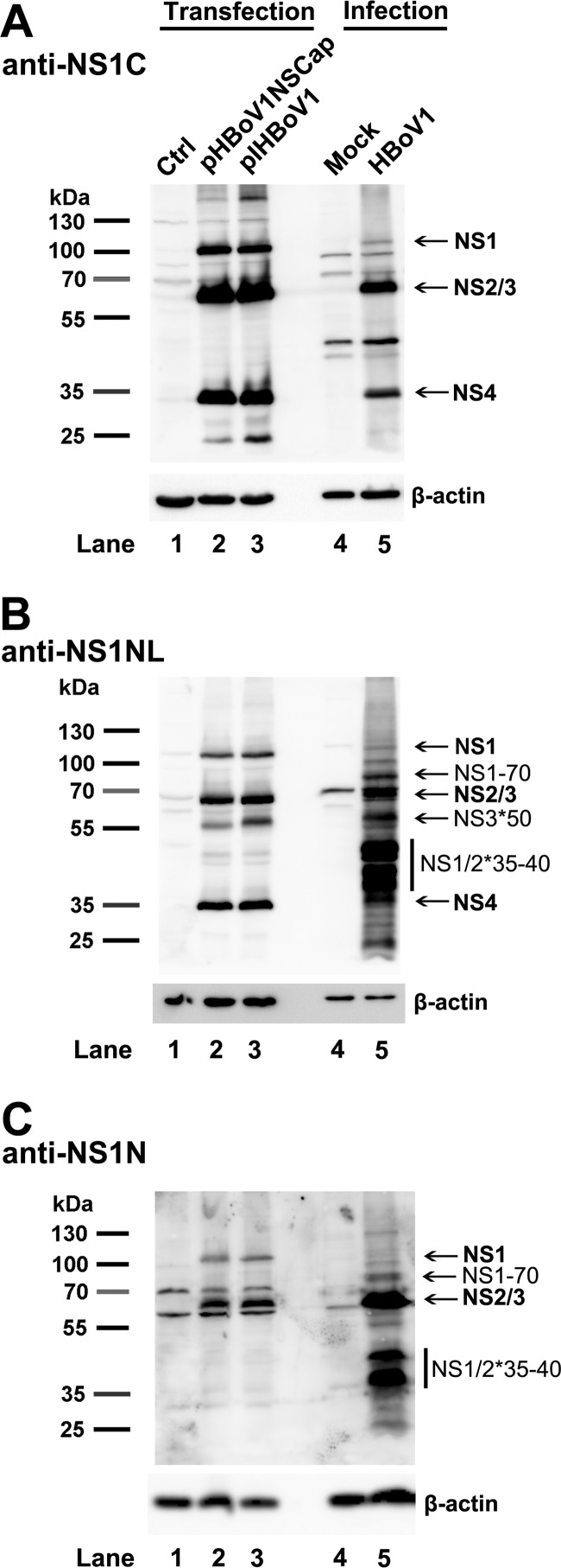
FIG 6.
Detection of HBoV1 NS2, NS3, and NS4 proteins in pIHBoV1-transfected HEK293 cells and HBoV1-infected HAE cells. HEK293 cells were transfected with pHBoV1NSCap or pIHBoV1. At 2 days posttransfection, cells were lysed. HAE-ALI cultures were infected with HBoV1 at an MOI of 10 (DRP/cell). At 14 days p.i., infected cells in the ALI cultures were lysed. The lysates of both transfected and infected cells, as indicated, were then analyzed by Western blotting using anti-NS1C (A), anti-NS1NL (B), and anti-NS1N (C) antibodies. The identities of detected proteins are shown with arrows at the right of the blot. Blots were reprobed for β-actin as a loading control.

