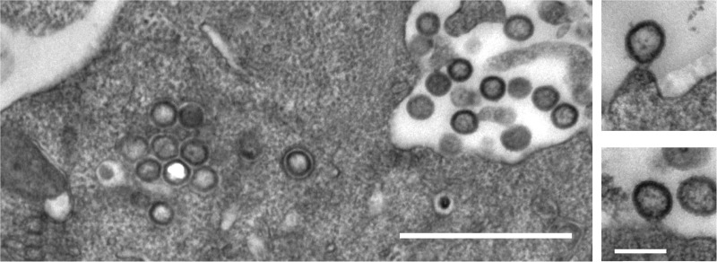
FIG 2.
Thin-section electron micrographs of HIVunc(LZ)-expressing cells. HEK293T cells were transfected with plasmid pCHIVunc(LZ), harvested at 36 h posttransfection, and fixed with 2.5% glutaraldehyde. Postfixation, embedment, and preparation for thin-section electron microscopy were performed as described previously (45). Bars, 1 μm (left) and 200 nm (right).

