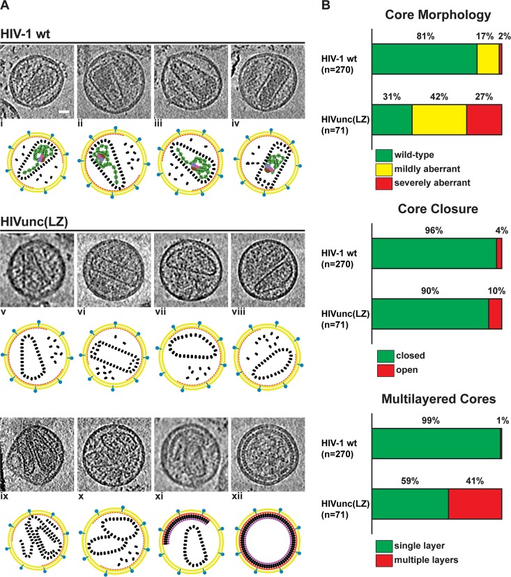
FIG 5.
Morphology of HIVunc(LZ) virions analyzed by cryo-electron tomography. (A) Gallery of computational slices through tomographic reconstructions showing a comparison between HIV-1 wt and HIVunc(LZ) particles. (i to iv) HIV-1 wt particles presenting typical conical and tubular cores encasing the condensed RNP density, as described in reference 27. (v to xi) HIVunc(LZ) mature particles showing the representative core morphologies: closed, single-layer, wt cores (v and vi); closed, single-layer, mildly aberrant cores (vii and viii); a closed, multilayer, wt core (ix); closed, single-layer multiple cores with a mildly and severely aberrant morphology (x); a closed, single-layer core in a particle also containing a partial immature lattice (xi). (xii) Immature HIVunc(LZ) particle. Bar, 20 nm. (B) Statistical analysis of the observed phenotypes.

