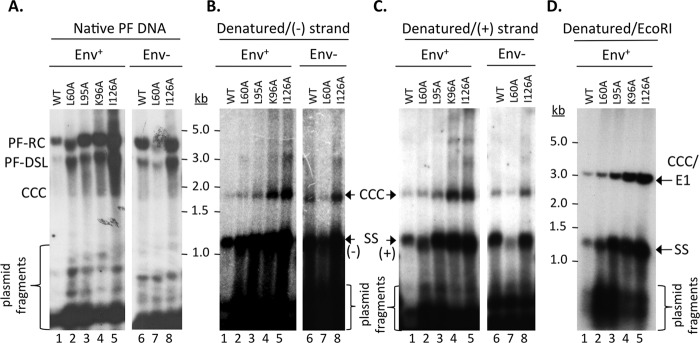
FIG 4.

Analysis of HBV PF DNA. HBV-replicating plasmids as in Fig. 1 were transfected into HepG2 cells. PF DNA was extracted and analyzed by Southern blotting, using an HBV-specific DNA probe (A and D) or a (−)-strand-specific (B) or (+)-strand-specific (C) RNA probe. Plasmid DNA copurifying with the PF HBV DNA was degraded by DpnI digestion as described in Materials and Methods. The plasmid fragments are indicated (A). Following DpnI digestion, the DNA was further denatured by boiling before analysis (B and C) or boiling followed by EcoRI digestion (D). SS, single-stranded [full-length (−)- or (+)-strand] DNA derived from the denatured PF-RC and PF-DSL DNA; CCC/E1, CCC DNA linearized by EcoRI digestion.
