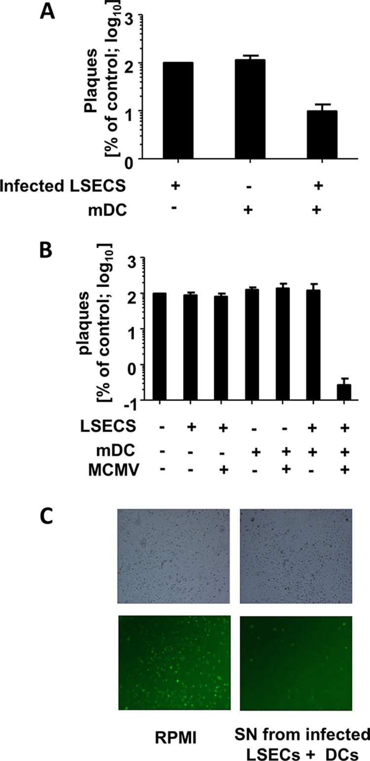
FIG 3.

Direct contact between CD11c+ mDC and infected target cells is required for secretion of a factor, which can block mCMV replication. (A) Virus growth in a two-chamber assay. LSEC were seeded in triplicates in the lower chamber and infected with MCMVr (MOI 0.1), while the upper chamber was seeded with mDC, infected LSEC (MOI 0.1), or mDC and infected LSEC. Virus titers were determined by plaque assay in supernatants from the lower chamber and normalized to values from the control group (no mDC in the upper chamber). Shown is the growth as a percentage of the control. Cells in the upper chambers are indicated below the x axis. Experiments were performed twice, and data were pooled. (B) Single cultures or cocultures of LSEC and mDC were infected (MOIs of 0.01 for LSEC and 1 for DC) or left uninfected. Supernatants were taken at day 6 or 7, filtered to prevent virus carryover, and transferred to fresh LSEC cultures infected with MCMVr (MOI of 0.1). Virus titers at day 6 postinfection were normalized to LSEC infected in the presence of standard cell culture medium (RPMI) and are shown on the y axis. Keys below the x axis indicate SN used as medium during second infection. Data from three independent experiments were pooled and are shown. (C) Representative bright-field and fluorescence microscopy pictures of LSEC infected at an MOI of 1 with MCMVr cultured in SN taken from coculture or medium control. Pictures were taken at 20 h postinfection.
