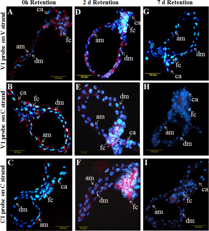
FIG 7.

TYLCV localization in midguts dissected from B. tabaci adults that had acquired TYLCV for 8 h and retained it on cotton plants for 0 h (A to C), 2 days (D to F), and 7 days (G to I). Localization assays were performed by using FISH with the following fluorescent probes: V1 for CP on the virus DNA strand (A, D, and G), cV1 for the CP gene sequence on the replicative DNA form of the virus (B, E, and H), and cC1 for the Rep gene sequence on the replicative DNA form of the virus (C, F, and I). All probes were conjugated to Cy3 dye. Blue indicates DAPI staining of nuclei. am, ascending midgut; dm, descending midgut; fc, filter chamber; ca, ceca.
