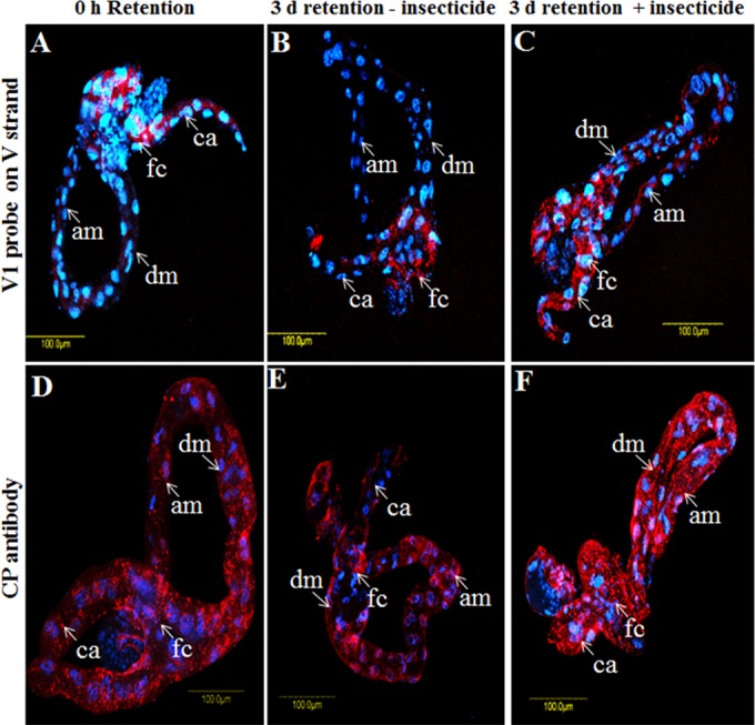
FIG 8.

TYLCV (red) localization in midguts dissected from B. tabaci B biotype adults under various treatment conditions. Shown are data from FISH (A to C) using the TYLCV V1 probe and immunolocalization analysis (D to F), using anti-TYLCV CP antibody after virus acquisition for 8 h (A and D) and retention for 3 days without acetamiprid treatment (B and E) or after acetamiprid treatment for 3 days (C and F). The blue signal is DAPI staining of nuclei.
