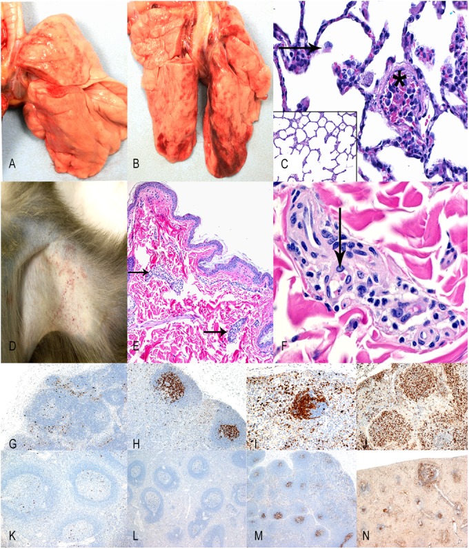
FIG 7.

Pathology. No gross changes in the lungs are evident from day 1 p.e. (A) and day 9 p.e. (B). (C) HE stain of the lungs on day 9 p.e. (magnification of ×200) shows mild increases in alveolar histiocytes (arrow) and fibrin thromboemboli within blood vessels (asterisk) compared with normal day 1 p.e. lung (inset; 100× HE). (D) Axillary haired skin with mild macular rash day 9 p.e. (E) HE stain (magnification of ×100) of day 9 p.e. axillary haired skin that contains superficial blood vessels with perivascular inflammation (arrows) and no hemorrhage in the dermis. (F) Higher magnification (400× HE) of superficial dermal blood vessel shows perivascular inflammation and fibrin within blood vessels (arrow). Caspase 3 IHC (magnification of ×100) staining of the tracheobronchial lymph node on days 3 (G), 5 (H), 7 (I), and 9 (J) p.e. illustrate the progressive and profound apoptosis of lymphocytes in the germinal centers of lymphoid follicles. Caspase 3 IHC staining (magnification of ×100) of the spleen on days 3 (K), 5 (L), and 7 (M) p.e. illustrates occasional to focal apoptosis in germinal centers of splenic corpuscles that progress to diffuse apoptosis on day 9 p.e. (N).
